A broad thin plate that forms the lateral part of the pterygoid process and gives attachment to the lateral pterygoid muscle on its lateral surface and to the medial pterygoid muscle on its medial surface. A variable pterygospinous process on its irregular posterior border is connected by a ligament sometimes ossified to the sphenoid spine 1.
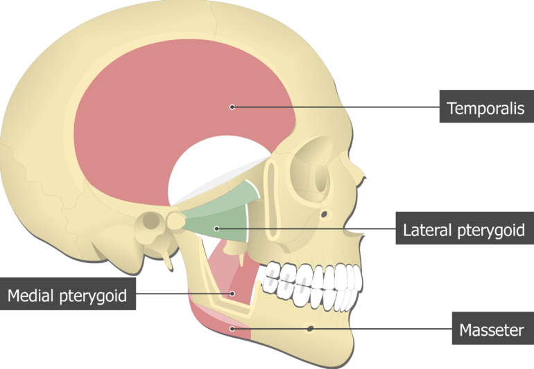
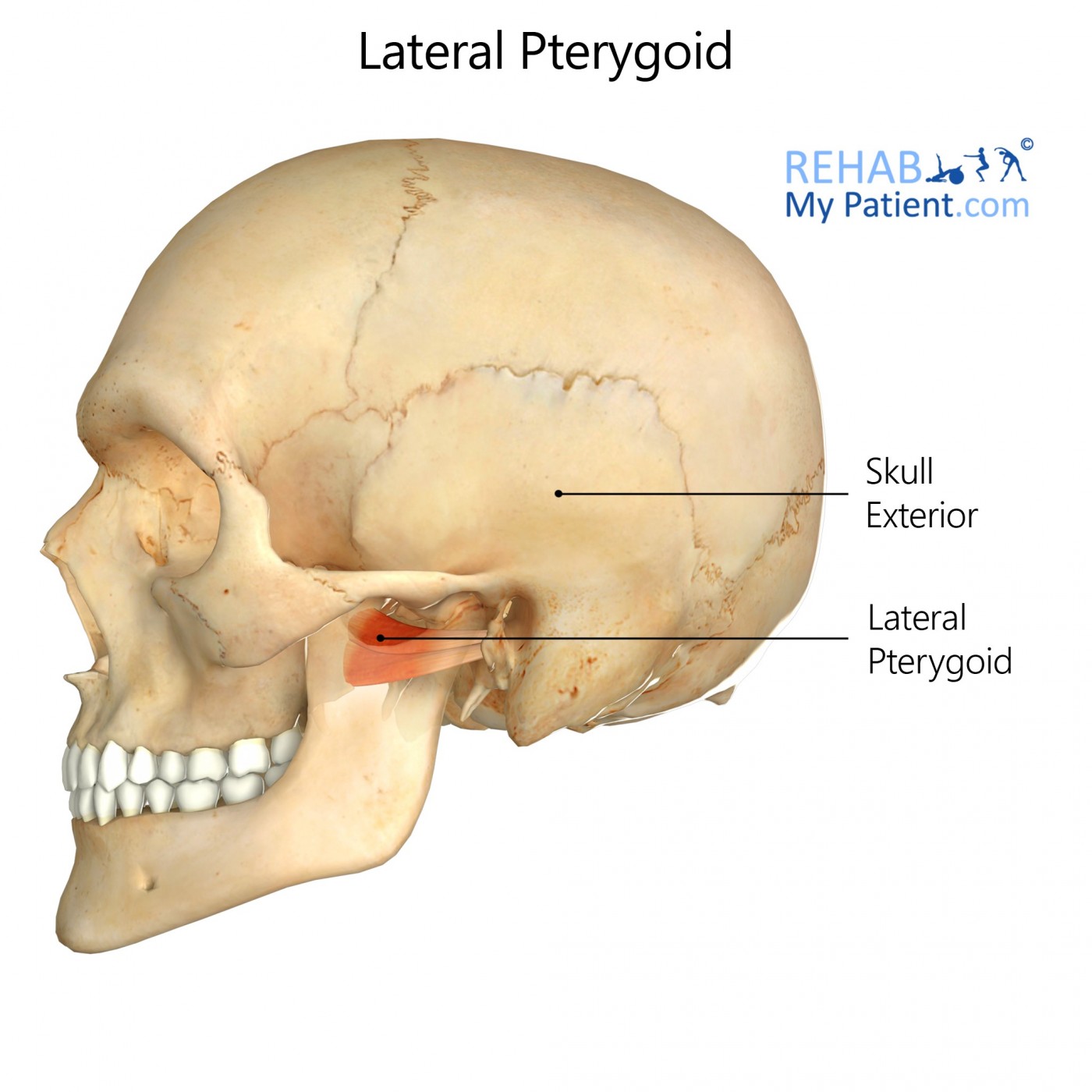
Lateral Pterygoid Muscle Attachments Actions Innervation
T he medial pterygoid muscle a major elevator of the jaw is a square-shaped masticatory muscle located on the medial aspect of the lower jaw bilaterally.
. The spaces separated the medial and lateral plate are pterygoid. The medial pterygoid muscle is a thick and square shaped muscle. Medial pterygoid muscles arise from the lateral pterygoid plates of the sphenoid bone and insert on the mandible.
PATTERN AND MANAGEMENT OF IATROGENIC DISPLACEMENT OF TEETH IN MAXILLOFACIAL ANATOMICAL SPACES. Tap card to see definition. The medial pterygoid plate is narrower and longer than the lateral.
The medial pterygoid plate or medial pterygoid lamina of the sphenoid is narrower and longer than the lateral pterygoid plate. Medial surface of lateral pterygoid plate sphenoid bone Pyr Lower and back part of medial surface of ramus and angle of ma Elevation mandible Clenches teeth Protrudes mandible Late. These movements maximize the efficiency of the teeth while chewing or grinding foods of various consistencies.
The suspected mechanism is due to force transduction through the medial and lateral pterygoid muscles when acute displacing force is placed on the mandible. The medial pterygoid along with the masseter allows the jaw to move in a vertical direction as it contracts and relaxes. Deep head medial side of lateral pterygoid plate and fossa between medial and lateral plates.
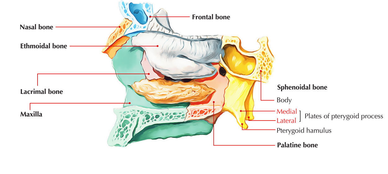
The medial pterygoid inserts on the pterygoid tuberosity of the mandible and medial inner surface of the mandibular ramus. A long narrow plate that forms the medial part of the pterygoid process terminates in the pterygoid hamulus and forms with its lateral. The lateral pterygoid muscle via its lower head separates the two distinct heads.
This muscle lies medial to the lateral pterygoid muscle. Each process consists of a medial pterygoid plate and a lateral pterygoid plate the latter of which serve as the origins of the medial and lateral pterygoid muscles. The lateral pterygoid allows the jaw to move in a horizontal direction during mastication.
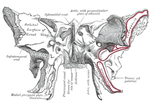
Each pterygoid process projects inferiorly from the junction of the body and greater wing of the sphenoid bone and bifurcates into a medial pterygoid plate and a lateral pterygoid plate. The medial pterygoid is a thick quadrilateral muscle. INSERTION Medial aspect of angle of mandible.
Aassits the lateral pterygoids in moving the jaw side-to-side. The pterygoid process is a thin plate of bone. Click card to see definition.
Medial pterygoid is a thick quadrilateral muscle that connects the mandible with maxilla sphenoid and palatine bones. Superior pharyngeal constrictor is attached towards inferior end of the medial pterygoid plate. Tuberosity of maxilla and pyramidal process of palatine bone.
It has a second slip of origin from the lateral surfaces of the pyramidal process of the palatine and tuberosity of the maxilla. Called also lateral pterygoid plate. Lateral pterygoid muscle has its the lower part connect to the lateral part of the lateral pterygoid plate.
The deep head is the major component and is attached to the medial aspect of the lateral pterygoid plate of the sphenoid bone. The smaller superficial head has its origin mainly from the maxillary tuberosity with some fibers arising from the pyramidal process of the palatine bone. This retrospective case series will review the relevant anatomy and propose a mechanism of isolate pterygoid plate fractures associated with mandible fractures.
The medial pterygoid along with the masseter allows the jaw to move in a vertical direction as it contracts and relaxes. At the inferior tip of the medial pterygoid plate is the small hook-shaped process the pterygoid hamulus. The medial pterygoid muscle has a superficial and a deep head which arise from different areas of the jaw.
Called also lateral pterygoid plate. The lateral pterygoid plate forms the medial margin while the maxilla forms the medial aspect of this space18 In the present study 4 impacted maxillary molars were pushed into infratemporal space during exodontias. -Belongs to the group of masticatory muscles along with the lateral pterygoid masseter and temporal muscles.
Isolated lateral pterygoid plate fractures ie. The lateral surfaceof this plate forms part of the pterygoid fossa the medial surface constitutes the lateral boundary of the choana or posterior aperture of the. The lateral surface of this plate forms part of the pterygoid fossa the medial surface constitutes.
The medial pterygoid plate is narrower and longer while the lateral pterygoid plate is broad thin and everted. -Consists of two heads. Moreover what artery supplies the medial and lateral pterygoid muscles.
The pterygoid hamulus is a hook-shaped bony process located bilaterally on each medial pterygoid plate of the sphenoid bone posterior and medial to each maxillary tuberosity. Both pterygoids protract the mandible and move it from side to side during chewing. It is also known as internal pterygoid muscle.
Medial pterygoid muscle consists of two heads. Without an associated medial pterygoid plate fracture or a Le Fort fracture associated with mandible fractures. Each process consists of a medial pterygoid plate and a lateral pterygoid plate the latter of which serve as the origins of the medial and lateral pterygoid muscles.
It arises from the medial surface of the lateral pterygoid plate and the grooved surface of the pyramidal process of the palatine bone. There is a long deep fossa internal pterygoid fossa between the pterygoid bone and the pterygoid process of the sphenoid bone. In the pterygoid process you will find a concave and smooth medial surface whereas the lateral surface is convex and possesses two parallel ridges.
It belongs to the group of masticatory muscles along with the lateral pterygoid masseter and temporal muscles. Pterygoid hamulus bursitisPterygoid hamular bursitis is a rare craniofacial pain syndrome used to describe palatal and pharyngeal pain due to an enlarged pterygoid hamulus. ACTION Elevates the mandible and assits in closing the jaw.
The medial pterygoid also elevates the mandible. The deep head of the muscle originates from the medial surface of the lateral pterygoid plate of the sphenoid bone. It curves lateralward at its lower extremity into a hook-like process the pterygoid hamulus around which the tendon of the Tensor veli palatini glides.
It has two heads of origin. The superficial head is attached to the maxillary tuberosity and the pyramidal process of the palatine bone. Although having different origins both heads insert on the inner.
In patients with identified isolated pterygoid plate factures a dedicated CT of the mandible may be indicated to assess for associated mandibular fracture even in patients whose clinical examinations have had. -Thick quadrilateral muscle that connects the mandible with maxilla sphenoid and palatine bones. Medial pterygoid muscle is attached towards medial part of the lateral pterygoid plate.
Origin and insertion of medial pterygoid by Anatomy Next Action. What muscle is medial to the pterygoid plate. It curves lateralward at its lower extremity into a hook-like process the pterygoid hamulus around which the tendon of the Tensor veli palatini glides.
A broad thin plate that forms the lateral part of the pterygoid process and gives attachment to the lateral pterygoid muscle on its lateral surface and to the medial pterygoid muscle on its medial surface. The lateral pterygoid allows the jaw to move in a horizontal direction during mastication.

Anatomy Trains Australia Nz Friday Anatomy Fact The Beautiful Pterygoids With The Coolest Name I Have A List Of Favourite Muscles And This One Is Top Of My List Along
Pterygoid Processes Of The Sphenoid Wikipedia
![]()
Medial And Lateral Pterygoid Muscle Anatomy And Function Kenhub
Lateral Pterygoid Rehab My Patient

Infratemporal Fossa Lo Pterygoid Plates Youtube

Lateral And Medial Pterygoid Diagram Quizlet
Pterygoid Processes Of The Sphenoid Wikipedia
Pterygoid Process Earth S Lab
0 comments
Post a Comment